
Proyect 018‐ABEL‐CM‐2013
Automated segmentation and disease progression analysis of focal brain pathology
using novel knowledge based algorithms
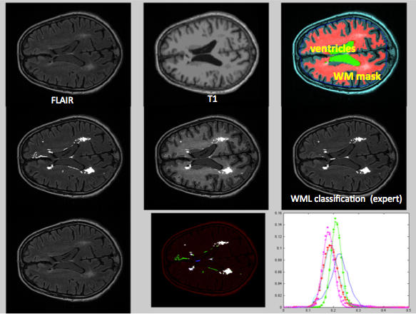
The presence of leukaraisosis or white matter lesions (WML) in the brain of elderly individuals is linked to increased risk of stroke, cognitive impairment, dementia and ultimately, death. Magnetic resonance imaging (MRI) is by far the most sensitive modality for detecting WMLs and MRI is consequently a very central diagnostic procedure in the elderly population. Manual WML segmentation is very time-consuming and prone to user-bias, which has resulted in several attempts to generating automated analysis tools for WML segmentation. In this project we will develop an automated WML quantification and analysis tool using available MRI data from large on-going prospective MR studies in patients with different neurodegenerative diseases. The tool will be developed using novel object based machine learning algorithms and preliminary results validated against radiological gold-standard indicate that the tool out-performs other available WML segmentation tools available currently. The algorithms developed as part of the project are also likely to have application for other types of focal brain lesions like multiple sclerosis and brain tumors
This project forms part of a large multi-national EU-funded project with the overall aim of identifying biomarkers predicting future development of dementia. Identification of premorbid (before disease manifestation and irreversible brain damage has occurred) biomarkers is considered crucial for development of effective future treatments of Alzheimer’s disease and other types of dementia. To this end, the availability of a robust and automated WML detection and characterization tools has been identified as a critical component for successful interpretation of MR findings. Given that manual WML segmentation is extremely time-consuming and operator-dependent, fully automated techniques are considered a prerequisite for practical use in large imaging studies. WML-features, as visualized on MRI are considered to contain extremely relevant information reflecting the pathogenesis of the disease, detailed WML characterization is therefore strongly desired as part of the overall image based disease phenotyping. The availability of a robust and automated WML detection and characterization tool would therefore contribute significantly to the advance of knowledge by providing user-independent and comprehensive analysis of multiple WML features and assessment of their correlation to other phenotypes and genotypes associated with the development of dementia. The developed methodology will also be extended for use in other focal brain pathologies with similar processing requirements including multiple sclerosis (MS) and primary brain tumors glioma in collaboration with on-going prospective studies at Oslo University Hospital (OUS).
Budget (in euros): 100,250.00 Euros
This Budget is dedicated to the coordinated mobility of researchers from UNED and IVS within the frame of this interinstitutional project.
Publications:
Rincón M, Díaz-López E, Selnes P, Vegge K, Altmann M, Fladby T, Bjørnerud A. Improved Automatic Segmentation of White Matter Hyperintensities in MRI Based on Multilevel Lesion Features. Neuroinformatics. 2017 Jul;15(3):231-245. doi: 10.1007/s12021-017-9328-y